FASTest® LEISH - Instructions for Use
Test-kit for the qualitative detection of antibodies against Leishmania infantum in whole blood, plasma or serum of the dog.
Vetlab Supplies
Specialists in Veterinary Laboratory Supplies
Supplied Exclusively To The UK Veterinary Market By Vetlab Supplies Ltd
Visit Our Website: www.vetlabsupplies.co.uk
Telephone: 01798 874567
Email: info@vetlabsupplies.co.uk
Manufacturer: DIAGNOSTIK MEGACOR
6912 Hörbranz - AUSTRIA
www.megacor.com
1. Information on the Test-Kit
Test-Kit Components
- 1 test-kit FASTest® LEISH contains:
- 2*, 10**, 25*** or 50**** test cassettes coated with Leishmania infantum antigens
- 1 dropper bottle A with *1.0 ml, **3.0 ml, ***7.5 ml or ****2×7.5 ml buffer diluent
- 2, 10, 25 or 50 disposable plastic pipettes
- 1 instructions for use
Stability and Storage
Store at 15-25°C. Expiry date: see label.
Application and Abbreviations
- For veterinary use only
- LOT: Lot number
- In vitro diagnosticum
- i Follow instructions for use precisely
- ! Do not use test-kit components from different kits, lot numbers or beyond stated expiry date.
- B - TEST line, C - CONTROL line, LF - Lateral flow
Liability
The entire risk due to the performance of this product is assumed by the purchaser. The manufacturer shall not be liable for indirect, special or consequential damages of any kind resulting from the use of this product.
Accuracy
- Sensitivity: 100%
- Specificity: 98%
- (Comparison Method: IFAT, ELISA)
2. Introduction
The visceral leishmaniosis of the dog is caused by the protozoon Leishmania infantum world-wide. Dogs and other canines are the reservoir for leishmaniasis in humans (zoonosis). To date leishmaniosis was known in Leishmania free regions as a pure travel or import disease. New investigations show increased sporadic occurring autochthonous cases of leishmaniosis in so far Leishmania free regions. The vectors, sandflies (Phlebotominae), admittedly need a subtropical to tropical climate, which however is not geographically dependent on such climatic zones. There are first scientific verified discoveries of sandflies in temperate zones. Furthermore, an infection via mating (urine/sperm), via diaplacentar transmission and via blood transfusion are discussed.
Leishmania are transferred by sand-flies via stings. They infest and reproduce in macrophages and cells of the reticuloendothelial system (among others liver, spleen, bone marrow, lymph nodes). Dependent on the Leishmania zymodeme and the immune status of the dog, there are variable clinical symptoms with dermatological (different skin and claw alterations) and visceral (apathy, fever, nose bleeding, lameness, kidney failure) manifestations.
Due to the individual extremely variable incubation times, from a few months to several years, infested animals can be free of symptoms during that time. The detection of Leishmania antibodies can be pointing at an initiating or an existing infection. Thus, suspected animals and animals from endemic leishmaniosis regions (travel or import) should be tested serologically for antibodies repeatedly in an interval of 2-4 weeks.
Animals from endemic areas and asymptomatic animals can show borderline to weak antibody titre ("seroprevalence"), whereas clinical diseased animals show a clear increase of titre between two tests ("disease prevalence"). Therefore the indirect detection of antibodies with FASTest® LEISH gets a greater diagnostic importance.
3. Information on the Specimen Material
Approximately 40-50 µl (1 drop of attached plastic pipette) 15-25°C warm whole blood (WB, with anticoagulant), plasma (P) or serum (S) are needed. Native blood without anticoagulant should not be used due to potential micro agglutination (e.g. migration delay on the membrane, unspecific reaction)! Mix the sample material well before use!
Non-cooled (15-25°C), WB, P and S should be tested within 4 hours! At 2-8°C, WB, P and S can be stored up to 4 days. Serum and/or plasma samples can be permanently stored at minimum -20°C.
Keep in mind that the sample material, as well as all used test-kit components, should have reached room temperature at the time of application.
Endogeneous and exogeneous interfering substances of the sample (e.g. albumin, fibrinogen, lipids, CRP, heterophilic antibodies, especially type IgA, as well as viscosity, pH-value and excess EDTA) as well as native blood can cause interferences (matrix effects) that can influence the target measurement. These can lead to an impaired LF and/or unspecific reactions on B and C.
4. Specimen Collection and Preparation
No specimen preparation necessary.
ATTENTION: Partially filled and/or insufficient mixed EDTA, Citrate or Heparin tubes could create invisible microclots resulting in lateral flow delay and/or unspecific reactions (e.g. greyish shadow like lines).
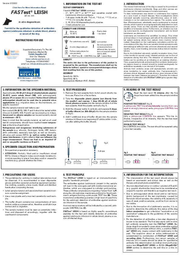
5. Test Procedure
- Remove the test cassette from its foil pouch shortly before use. Place it on a flat surface.
- Take the disposable plastic pipette (not directly from the needle!) and express 1 drop (40-50 µl) of whole blood, plasma or serum into the sample window A of the test cassette. Hold the pipette vertically (fig. 1).
- Hold the dropper bottle A vertically and express 5 drops of buffer diluent (ca. 200-250 µl) into the sample window A (fig.2).
- Add 1 additional drop of buffer diluent into the sample window A if there is no beginning LF visible within 1 minute after adding the buffer diluent.
fig. 1
fig. 2
5 drops
6. Reading of the Test Result
Read the test result 15 minutes after the five drops have been added into the sample window A.
Positive Test Result (fig. 3)
A pink-purple TEST line of any intensity (varying from very weak to strongly intensive) and a pink-purple CONTROL line appear.
Negative Test Result (fig. 4)
Only a pink-purple CONTROL line appears. This line indicates, irrespective of its intensity, that the test has been performed properly.
Invalid Test Result
No CONTROL line visible. The test should be repeated using a new test cassette.
fig. 3 Positive Test Result
fig. 4 Negative Test Result
B C
7. Precautions for Users
The guidelines for working in medical laboratories must be observed. It is recommended to wear disposable gloves and other personal protective equipment (protective clothing, possibly a face mask). Wash and disinfect hands after completing the test.
Label sample material and associated test cassette to ensure a precise assignment.
Use a new pipette and a new test cassette for each sample.
The buffer diluent contains low concentrations of toxic sodium azide as a preservative, therefore avoid skin/eye contact and/or ingestion.
The sample material must be seen as potentially infectious and disposed of accordingly, together with the used test-kit components.
8. Test Principle
The FASTest® LEISH is based on an immunochromatographic "sandwich principle".
The antibodies against Leishmania present in the sample will react in the conjugate pad with mobile monoclonal antibodies, which are conjugated to colloidal gold particles. These antibody complexes are migrating ("lateral flow", LF) along the nitrocellulose membrane and bind to fixed Leishmania antigens forming a pink-purple TEST line (B). These monoclonal antibodies guarantee a high level of specificity for the aetiologic detection of antibodies against Leishmania infantum in the sample.
A correct test procedure will be indicated by a second, pink-purple CONTROL line (C).
FASTest® LEISH is based on highly specific recombinant peptides for the fast and reliable detection of antibodies against Leishmania infantum in whole blood, plasma or serum of infected dogs.
9. Information for the Interpretation
The interpretation of the test result should always be based on anamnestic and clinical data as well as the therapy and prophylaxis possibilities.
Any non-described colour or contour variation of B and C (e.g. greyish, shadow-like lines) has to be considered as unspecific reaction and therefore as negative test result.
Due to anticoagulated whole blood and/or red hemoglobin background of the test membrane, caused by hemolytic blood samples, the visibility of B, especially in case of weak positive samples, could be from worse to not visible.
Due to the innovation of a Leishmania vaccine, it is required to determine the antibody titre status of the dog before vaccination to get a decision "vaccination or no vaccination" adequate to the guidelines of the vaccine manufacturer.
For the detection of antibodies, a two-step diagnosis is known to be standard. The first step starts with in-clinic IgG antibody screening test like FASTest® LEISH. Because dogs from endemic areas show antibodies against Leishmania on principle without clinic, a positive FASTest® LEISH only means contact with Leishmania in the past. The suspicion about an active leishmaniosis is substantiated by combination of FASTest® LEISH and according clinic. Furthermore, two serum samples at intervals of 2-4 weeks should be taken for quantitative antibody titre determination via indirect immunofluorescence test (MegaFLUO® LEISH) or ELISA (MegaELISA® LEISH canine) to determine the end titre or a titre increase.