1. Introduction
The CONTEC CMS1600B-vet is a handheld dual-probe color Doppler ultrasound diagnostic system. It consists of a main unit and dedicated control software. This system utilizes ultrasonic Doppler technology and ultrasonic echo theory to gather information on blood flow movement, tissue movement, and to generate images of organ tissues. Its compact size and ease of operation make it suitable for clinical ultrasound examinations in various animals, including canine, feline, ovine, bovine, and equine species.
2. Product Overview
The CMS1600B-vet system is designed for veterinary use, offering a range of adjustable parameters to suit different diagnostic needs. Key features include:
- Dual-Probe Design: Integrates both Convex and Linear probes for versatile applications.
- Wireless Connectivity: Effective WiFi transmission distance of not less than 10 meters (in open areas) between the main unit and control software.
- Rechargeable Battery: Supports wireless charging for convenience.
- Display Features: Full-screen display support, image flip function, and cine-loop function.
- Working Modes: B, 2B, B/M, C, PW.
- Image Adjustment: 8-segment TGC (Time Gain Compensation) adjustment.
The device is compact and ergonomically designed for handheld use, as shown in the image below.

Figure 2.1: The CONTEC CMS1600B-vet handheld ultrasound device.
The system includes the main ultrasound unit, a charging cable, and a software disc, all packaged for safe transport and storage.
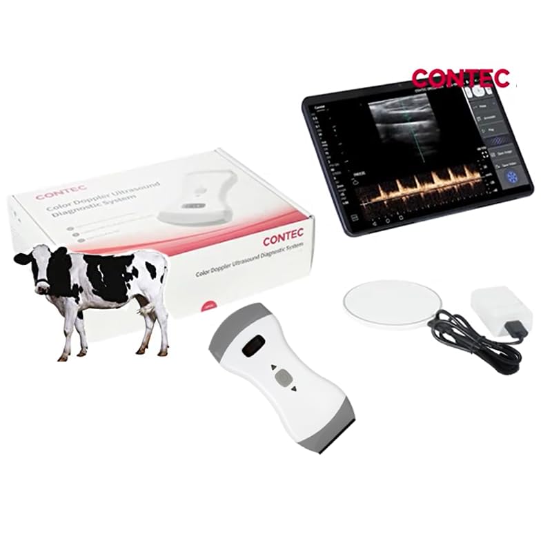
Figure 2.2: Product packaging and included components.
3. Setup
3.1 Charging the Device
Before initial use, ensure the device is fully charged. The CMS1600B-vet supports wireless charging. Connect the provided charging cable to a suitable power source and place the device on the charging pad, or directly connect the USB-C cable to the device's charging port.

Figure 3.1: Connecting the charging cable to the device.
3.2 Software Installation and Connection
The CMS1600B-vet operates with specific control software. Install the software on your mobile phone, tablet, or computer. The system is compatible with both iOS and Android operating systems.

Figure 3.2: The ultrasound software running on a smartphone and tablet, compatible with Android and iOS.
Once the software is installed, power on the ultrasound device. The device will broadcast a WiFi signal. Connect your mobile device or computer to this WiFi network. The effective transmission distance for the WiFi connection is approximately 10 meters in an open area.

Figure 3.3: Wireless connection of the ultrasound device to a tablet via WLAN.
4. Operating Instructions
4.1 Powering On/Off
Press and hold the power button located on the device to turn it on or off. The device features an indicator light and a small display to show battery status and operational mode.

Figure 4.1: The device in hand, showing the power button and battery indicator.
4.2 Performing an Examination
Once connected to the control software, select the appropriate probe (Convex or Linear) and working mode (B, 2B, B/M, C, PW) for the examination. Adjust parameters such as TGC (Time Gain Compensation) as needed for optimal image quality. The software supports full-screen display, image flip, and cine-loop functions for comprehensive analysis.

Figure 4.2: The ultrasound device being used, with the live image displayed on a connected tablet.
5. Clinical Applications
The dual-probe design of the CMS1600B-vet allows for a wide range of clinical applications in veterinary diagnostics:
- Linear Probe Applications:
- Vascular
- MSK (Musculoskeletal)
- Pediatrics
- Breast
- Carotid
- Thyroid
- Small Parts
- Convex Probe Applications:
- Abdomen
- Cardiac
- Gynecology
- Obstetric
- Urology
- Kidney
This versatility makes the system suitable for comprehensive diagnostic imaging across various animal types and medical conditions.

Figure 5.1: Overview of clinical applications for the Linear and Convex probes.
The integrated dual probe design is clearly visible, catering to different examination requirements for animals like cats and dogs.

Figure 5.2: The CONTEC CMS1600B-vet device alongside a kitten and a puppy, highlighting its application in veterinary settings.
6. Maintenance
6.1 Cleaning
Regularly clean the device, especially the probe surfaces, with a soft cloth dampened with a mild disinfectant solution. Ensure no liquid enters the device openings. Do not use abrasive cleaners or solvents.
6.2 Battery Care
To prolong battery life, avoid fully discharging the device frequently. Charge the device regularly, even if not in constant use. Store the device in a cool, dry place when not in use for extended periods.
7. Troubleshooting
- Device not powering on: Ensure the battery is charged. Connect to the charger and attempt to power on again.
- No image on screen: Verify that the device is powered on and successfully connected to the control software via WiFi. Check the WiFi connection status on your mobile device or computer.
- Poor image quality: Adjust the TGC settings in the software. Ensure proper contact between the probe and the animal's skin, using ultrasound gel as necessary.
- WiFi connection issues: Ensure your mobile device is within the 10-meter effective range of the ultrasound unit. Check for interference from other wireless devices.
8. Specifications
- Model: CMS1600B-vet
- Product Dimensions: 6.34 x 2.84 x 1.18 inches
- Weight: 1.41 Pounds
- Working Modes: B, 2B, B/M, C, PW
- TGC Adjustment: 8-segment
- Connectivity: Wireless (WiFi), effective range > 10m (open area)
- Charging: Wireless, USB-C
- Display Features: Full-screen display, image flip, cine-loop
- Compatibility: iOS, Android
- Applications: Canine, feline, ovine, bovine, equine, etc.
9. Warranty and Support
For detailed warranty information and technical support, please refer to the official CONTEC website or contact your authorized dealer. Keep your purchase receipt for warranty claims.


