Verlag Unser Wissen 6204179519
Decalcification in Histology User Manual
Published by Verlag Unser Wissen
Model: ISBN 6204179519

This image displays the front cover of the manual, featuring the title, authors, and a scientific illustration of a microscope with laboratory beakers, symbolizing the histological context.
1. Introduction to Decalcification in Histology
Demineralization is a rapidly growing and challenging aspect across various scientific disciplines, including astrobiology, paleoclimatology, geomedicine, archaeology, geobiology, dentistry, histology, and biotechnology. This manual provides comprehensive insights into the process of decalcification, particularly its application and significance within the field of histology.
Biomineralized structures and tissues are complex composites, naturally formed with an organic matrix and amorphous or crystalline minerals at the nano- or micro-scale. In human anatomy, the head and neck region presents a particularly intricate structure comprising both soft and hard tissues. While soft tissues generally pose fewer challenges for histochemical techniques, lesions affecting hard tissues necessitate a sophisticated and technically sensitive methodology for accurate interpretation and diagnosis.
2. Factors Influencing Decalcification
The selection of an appropriate decalcifying agent is critical and is influenced by four interdependent factors:
- Urgency of the Case: The speed at which decalcification needs to occur can dictate the choice of agent.
- Degree of Mineralization: The density and extent of mineral content in the tissue will affect the required strength and duration of the decalcification process.
- Scope of Investigation: The specific research or diagnostic goals will influence the method, as some techniques may preserve certain cellular components better than others.
- Required Staining Techniques: The chosen decalcifying agent must not interfere with subsequent staining procedures, which are crucial for microscopic examination.
3. Characteristics of an Ideal Decalcifying Agent
An ideal decalcifying agent should possess several key properties to ensure optimal results in histological preparation:
- It should ensure the complete removal of calcium from the tissue.
- It should cause minimal damage to cells and tissues.
- It must not interfere with subsequent staining processes.
- It should decalcify at an appropriate and controllable speed.
4. Setup and Preparation for Decalcification
Proper setup is crucial for effective decalcification. This involves preparing the tissue samples, selecting the appropriate decalcifying solution, and ensuring a suitable laboratory environment.
4.1 Tissue Sample Preparation
Before decalcification, tissue samples must be adequately fixed to preserve cellular morphology. Ensure samples are cut to an appropriate size to allow for efficient penetration of the decalcifying agent. Smaller, thinner samples generally decalcify faster and more uniformly.
4.2 Reagent Selection and Handling
Choose a decalcifying agent based on the factors discussed in Section 2. Common agents include strong acids (e.g., nitric acid, hydrochloric acid), weak acids (e.g., formic acid), chelating agents (e.g., EDTA), and electrolytic methods. Always handle these reagents with appropriate personal protective equipment (PPE) in a well-ventilated area or fume hood.
5. Operating the Decalcification Process
The decalcification process involves immersing the fixed tissue in the chosen decalcifying solution. The duration of immersion depends on the type of tissue, its size, and the strength of the decalcifying agent.
5.1 Monitoring Decalcification
Regularly monitor the progress of decalcification. This can be done through various methods, including:
- Physical Examination: Gently bending the tissue to check for flexibility.
- Chemical Test: Testing the decalcifying solution for the presence of calcium ions (e.g., using ammonium oxalate).
- Radiography: X-ray imaging can precisely determine the extent of decalcification, especially for larger or denser specimens.
Once decalcification is complete, thoroughly wash the tissue to remove residual decalcifying agent, which could interfere with subsequent processing steps.
6. Maintenance and Post-Decalcification Handling
Proper maintenance of samples and reagents ensures the integrity of the tissue and the accuracy of results.
6.1 Reagent Storage
Store decalcifying agents according to manufacturer guidelines, typically in cool, dark places, and in appropriate containers to maintain their efficacy and safety.
6.2 Tissue Storage
After decalcification and washing, tissues should be processed promptly for dehydration, clearing, and embedding. If immediate processing is not possible, store tissues in a suitable holding solution, such as 70% ethanol, to prevent degradation.
7. Troubleshooting Common Issues
Several issues can arise during the decalcification process. Understanding these can help in achieving optimal results.
7.1 Incomplete Decalcification
If tissue remains hard after the expected decalcification period, it may indicate insufficient time, an exhausted decalcifying agent, or too large a sample. Consider extending the decalcification time, replacing the solution, or re-sectioning the tissue.
7.2 Tissue Damage
Over-decalcification, especially with strong acids, can lead to tissue maceration, swelling, or loss of cellular detail, which compromises subsequent staining. Monitor the process closely and use milder agents or shorter durations for delicate tissues.
7.3 Staining Interference
Residual decalcifying agents can inhibit or alter staining reactions. Ensure thorough washing of tissues after decalcification to prevent this. Some agents may also permanently affect tissue affinity for certain dyes.
8. Book Specifications
This manual is presented in a paperback format, providing a comprehensive guide to decalcification in histology.
| Attribute | Detail |
|---|---|
| Publisher | Verlag Unser Wissen |
| Publication Date | October 24, 2021 |
| Language | German |
| Print Length | 56 pages |
| ISBN-10 | 6204179519 |
| ISBN-13 | 978-6204179513 |
| Item Weight | 3.25 ounces |
| Dimensions | 5.91 x 0.13 x 8.66 inches |
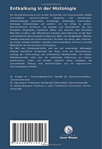
This image shows the back cover of the book, which includes a barcode with the ISBN-13 (9786204179513) and a summary of the book's content in German, along with author affiliations.
9. Authors and Contributors
This comprehensive manual is authored by:
- Sheeba Ali
- Puja Bansal
- Deepak Bhargava
Their expertise in the field of histology and related scientific disciplines has contributed to the detailed and informative content of this publication.
10. Further Reading and Support
For further academic inquiries or to explore related publications, please refer to the publisher's official channels. As this is a scientific publication, direct product support or warranty information is not applicable in the traditional sense. However, academic support regarding the content may be sought through the publisher, Verlag Unser Wissen.
For more information about this book or to purchase, please visit the product page: Amazon Product Page
Ask a question about this manual
Ask about setup, troubleshooting, compatibility, parts, safety, or missing instructions. Manuals+ will review the question and use this page’s manual context to help answer it.